Editorial
Managing micropthalmia in children
Published
4 years agoon
By
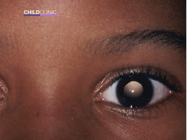
Micropthalmia is a severe development defect of the eye where one or both eyes are unusually small and have anatomic malformations. A gene mutation or certain types of drugs during pregnancy, which include isotretinoin (accutane) and thalidomide, are known causes of the condition.
A US Centre for Disease Control funded research estimates that about one in every 5,200 babies is born with micropthalmia. This data also suggests one of the risk factors of this condition includes maternal age of over 40, multiple births, infants of low birth weight and low gestational age.
To diagnose micropthalmia, a series of evaluations and examinations are conducted. They include:
Imaging
Micropthalmia is conspicuous and thus can be spotted quite easily after childbirth. If you notice the volume of your newborns’ eyes are smaller than normal, it is highly recommended to visit a pediatrician. Eye measurements are the first action in the diagnosis procedure. Ultrasound is usually first used to determine the length of the globe in the eyes of a child with micropthalmia and also examine their orbits.
After that, Magnetic Resonance Imaging (MRI) and electrophysiological tests are done. MRIs are useful for orbital evaluation and are used to locate orbital cysts. Electrophysiological tests on the other hand assess the severity of visual impairment and the level of abnormality. If the case is severe, visual evoked potential is used to determine whether any visual function is present. It also helps detect optical nerve dysfunction. The visual evoked potential test is used in conjunction with electroretinogram to identify if there is retinal dysfunction.
Family history
Since micropthalmia is mostly caused by a gene mutation, examination of other family members for similar ocular defects is important. This means testing for everything from micropthalmia, glaucoma, anterior segment malformation to optic nerve hypoplasia. These tests are conducted to provide a clue to a likely diagnosis or find an inheritance pattern.
Comprehensive anatomical evaluation
Micropthalmia can at times be associated with non-ocular anomalies. That means this ocular defect can come about as a result of a non-ocular syndrome. Some of the syndromes associated with micropthalmia include CHARGE, Goltz and Lowe syndrome. As a result of this relation, physical examination including a dysmorphology examination is paramount. This is also crucial because the syndromes that affect ocular development can also affect brain development.
Treatment options
Treatment is usually started early to give the children an improved chance of getting the best results. This is because the eye globe triples in volume between birth and adolescence, therefore providing the perfect window for varied solutions. It is, however, important to note that there is no way to create a new eye or restore complete vision. During the process of treatment, it is recommended that an ophthalmologist, an ocularist and an oculoplastic surgeon work together to maximise on the child’s visual potential. Some of the things they can do include:
Expansion of the eye socket
This is done in a specialist unit by an ocularist and oculoplastic surgeon who fit the child with a conformer (prosthetic eye that’s not painted). To avoid any facial deformity, this is recommended especially at an early age. Facial deformity comes about because reduction in ocular volume consequentially affects facial and orbital development, therefore causing underdevelopment of parts such as the bony orbit, eyelids and fornices. If unattended to, an underdeveloped socket also ruins the chance to wear prosthetics in later life. The condition is therefore treated early to ensure the cosmic deformities that would otherwise plague the child are curbed.
Medical therapy
In case there is detectable retinal function, the eyes are refracted and the treatment of any underlying amblyopia (partial or complete loss of vision in one eye) is made a priority. Glasses are often recommended as the lenses control the refraction and correct any resulting errors.
Development assessment
A child with severe micropthalmia often has no light perception. This often leads to unusual sleep patterns. Growth and development assessments can thus can be used to detect such abnormalities early. Melatonin supplements are usually prescribed to facilitate a normal sleep pattern.
This article was first published in the March issue of Parents Magazine