Love your ultrasounds (part 1)

An ultrasound is a chance to “meet” your baby.
The use of ultrasound makes it possible to see the womb using sound waves. Although ultrasound scanning is considered to be safe, some mothers are still adamant on having one. This is because they feel there is no enough long-term evidence of safety. Therefore, it should be done once a problem has been detected and a diagnostic ultrasound is required.
There are many times ultrasound scans gives valuable information to both you and your doctor. Information that will clearly improve the outcome of your pregnancy. Here is why this scan is important and why you should love it.
How it is conducted
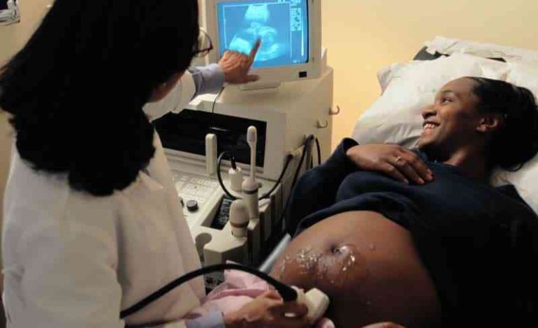
A trans-abdominal ultrasound in painless. A mother is usually required to take plenty of fluids before embarking on it for purposes of keeping her bladder full. In early pregnancy, this will help in pushing the uterus out of the pelvic cavity.
The gel that is applied on her belly helps with conduction of sound waves.
A transducer is then moved back and forth the mother’s abdomen, picking up the sound waves and feeding them on a computer, which displays them as image on the screen.
Compared to earlier, the development of the ultrasound scans has come a long way. Instead of looking at images that are difficult to interpret due to clarity, ultrasounds today can show your baby’s face in great detail.
Below are methods which use different imaging centers that perform the ultrasounds
A-mode – This is the simplest method of performing an ultrasound scan
B-mode / 2D mode- It is a two- dimensional way of viewing the foetus
3D ultrasound- It is the most commonly method used by obstetricians. It provides a 3D picture of a growing baby
4D ultrasound- It is similar to the 3D but provides a more realistic picture of your baby.


